


GENORAY MAKES INVISIBLE VISIBLE



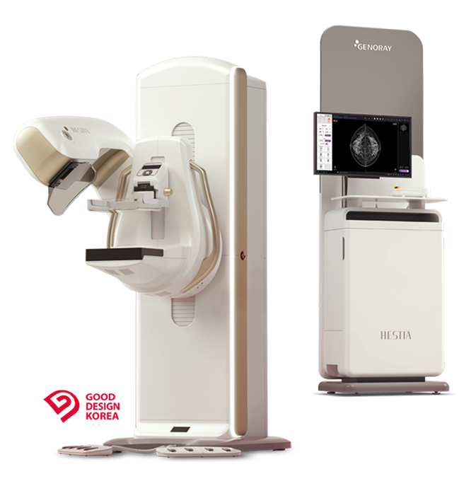



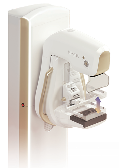
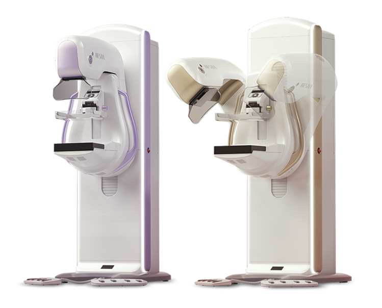
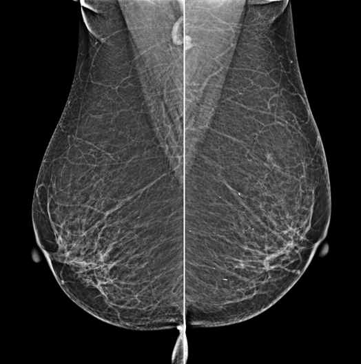
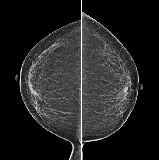
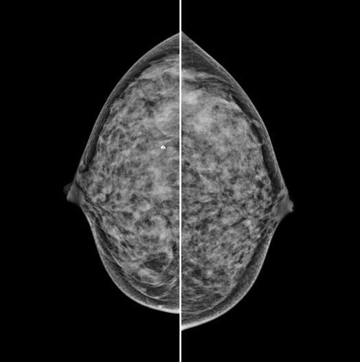
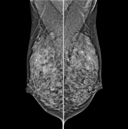
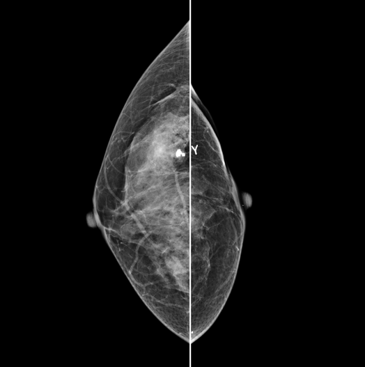
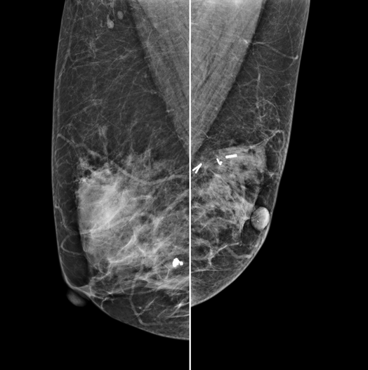
The X-ray tube rotates around the breast and takes high-definition, high-resolution images with high contrast from various angles.The images are reconstructed in 3D to provide accurate information about the location and condition of the lesion.
HESTIA supports "Synthetic 2D" that generates 2D images only by tomography shooting.
HESTIA tells Examinee the current status of diagnosis by voice guide and instruction screen.
To ensure patient safety, HESTIA provides and manages an automatic compression-release function.
HESTIA's LED lights gives its status in color to the operator in real time.

Emergency Stop Switch
For the examinee's safety, there is
"Emergency Stop Switch" on the operation panel.
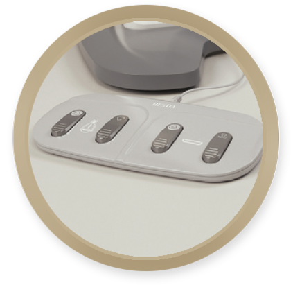
4-Button Foot Switch
HESITA is designed to allow for intuitive breast
compression and up-and-down movement of the gantry.
The L (Large) type that could only take 2D pictures can be upgraded into the T (Tomo) type that can also take 3D shots, It can be done with a simple software installation without additional H/W installation or system replacement.
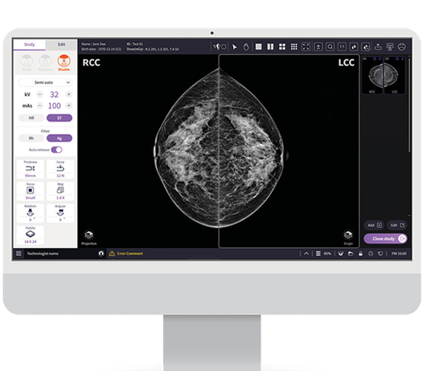
- Viewer optimized for Mammography's clinical
workflow
- Optimized image Acquisition & processing Workstation for HESTIA
- Supports DICOM 3.0 and realizes PACS linkage
- For the operator's convenience, HESTIA adopted Intuitive, highly visible user interface

Corporate Name GENORAY Co., Ltd. Business Registration No. 215-86-06482
CEO ByungUk Park
Global HQ 9F, Dongil Tower, 38 Teheran-ro 114-gil, Gangnam-gu, Seoul
Seongnam Plant 1 60, Dunchon-daero 541beon-gil, Jungwon-gu, Seongnam-si, Gyeonggi-do
Tel +82-2-2206-2200 Fax +82-2-2206-2299
